Forensic Imaging
The Forensic Imaging Bureau (FIB) houses some of the most sophisticated and high-tech photography equipment in the world. The 2,713-square-foot state-of-the-art facility contains a digital mini-lab, studio, computer lab, video editing room, and high-speed gun range. Through the use of these facilities and equipment, vital evidence can be documented. This helps medical examiners determine the cause of death in cases under investigation.
Leonard Wolf, CFPH, supervisor of the FIB, has been a photographer since 1984. He became a certified forensic photographer with the International Association for Identification (IAI) in 2006. There are four full-time staff photographers and up to three interns. Collectively they take more than 10,000 photographs per month, using a wide range of equipment.
Since 2006 almost all photographs taken have been with the use of digital cameras. A set of standard operating procedures insures the capturing and processing of digital images in a way that courts will accept.
The FIB’s computer lab uses some of the latest software to help document and present evidence. Here are some examples:
- Still images can be pulled from a surveillance tape for enhancement.
- 3-D images can be constructed to help determine the path of a bullet or a knife.
- A composite overlay of a boot print can be matched against a boot print on the skin of a victim who has been stomped by an assailant.
We are the only medical examiner department in the United States with its own indoor range that enables high-speed still and motion photography. Our digital video motion picture camera is capable of capturing 160,000 pictures per second. Our micro flash, designed for still photographs, has a flash duration of one-half millionth of a second.
Within the indoor range, tests with the weapon used in a crime can take place in a controlled environment. Results of these tests help demonstrate how close the victim may have been to the shooter, different gas dispersal patterns for different guns and other important findings.
The FIB’s digital mini-lab can produce color or black and white prints, scan almost all formats of film, and convert the images to digital media. It can process, print, and archive almost all formats of digital media. All digital images are processed and archived on a stand-alone and secure workstation and server.
Images that are too small to be seen with the naked eye can be recorded with the use of photo-microscopy and photomicrography. Utilizing this specialized technology, detailed, high-powered images of cross-sectioned tissue, gun cartridge casings or fibers can be created.
An area of increasing interest involves the taking of photographs with light other than full-spectrum white light. This includes the use of ultraviolet and infrared lights and filters, a modified digital camera, and a device called a Spex Light. With the use of these special techniques and equipment, conditions such as surface bruising or deep tissue bruising, and trace evidence, such as semen or blood, can be better detected.
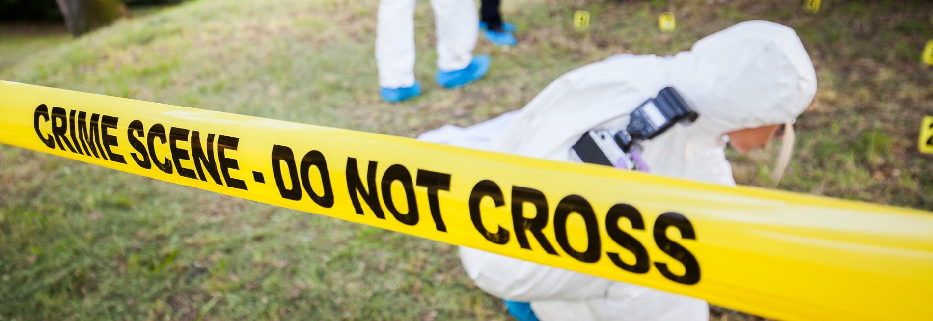
The FIB staff offers its services not only to doctors but also to various law enforcement agencies. We know and understand that photography plays a major role in the preservation of events. Photographs are often the most accurate form of documentation. Thus the camera becomes one of the most important tools in investigations, because it captures an image clearly and permanently.
Leonard Wolf
Forensic Imaging Bureau Supervisor
305-5452496
[email protected]
FORENSIC SPECIALTIES
Medical Examiner
Dr Benjamin Mathis, M.D.
Medical Examiner Building
1851 NW 10th Ave,
Miami, FL 33136
305-545-2400